Blepharoplasty Anatomy Explained | Upper and Lower Eyelid Structure
Understanding the Anatomy of the Eyelids with Dr. Balikian
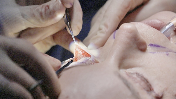
Blepharoplasty is often described as “eyelid surgery,” but the truth is more precise. It is surgery on one of the most delicate, layered and functionally important structures on the face.
A few millimeters of skin or fat in the wrong direction can change not only how the eyes look but how they blink, hydrate and protect the globe.

Understanding eyelid anatomy is the key to achieving natural, safe and lasting results.
This page breaks down how the upper and lower eyelids are built, how they age and why successful blepharoplasty depends on respecting these fine structures.
A separate page can explain how the procedure is actually performed, but everything starts with anatomy.
The eyelids control vision, eye protection, tear distribution and facial expression. They contain some of the thinnest skin in the body and a coordinated system of muscles, ligaments, fat pads and nerves.
Aging affects each layer differently:
Research shows that these combined changes can affect both appearance and function. Patients with dermatochalasis often experience reduced contrast sensitivity, obstruction of the superior visual field and increased forehead muscle activity, which improves after blepharoplasty.
Good blepharoplasty does not “change” your eyes. It restores the natural relationships between these structures.


The upper eyelid functions like a finely balanced shutter. Every blink requires coordinated movement of multiple layers, working together across a curved surface to protect the eye and maintain natural expression.

A clear understanding of these layers is essential, as each one plays a role in both appearance and function.
The anatomy, from superficial to deep, includes:
The eyelid contains the thinnest skin on the human body, making it especially prone to stretching and folding over time. Redundant skin is the hallmark of dermatochalasis and often contributes to heaviness and hooding.
This circular muscle allows for gentle blinking and firm eyelid closure. It also plays an important role in tear distribution and contributes to the natural contour of the eyelid.
Modern techniques emphasize preserving the orbicularis muscle to maintain normal blinking and avoid hollowing that can result from overly aggressive removal.
The orbital septum is a thin fibrous barrier that helps contain orbital fat. With aging, this layer can weaken, allowing fat to shift forward and create fullness or hooding, often referred to as pseudoherniation.
The strength and behavior of the septum can vary between individuals, influencing how these changes appear.
The central and nasal fat pads act as natural cushions and contribute to the soft fullness of a youthful upper eyelid. Over-removal can lead to a hollow or skeletonized appearance, which is more difficult to correct.
Preservation or careful repositioning is often preferred to maintain natural contour.
This tendon connects the levator muscle to the eyelid and is responsible for lifting the upper lid. Its attachments to the skin form the eyelid crease.
When the levator aponeurosis stretches or detaches, a condition known as ptosis can occur. In these cases, removing skin alone does not correct the underlying issue, and the muscle must be addressed to restore proper eyelid position.
These deeper structures contribute to eyelid tone, stability, and smooth movement. They help maintain eyelid shape, protect the cornea, and support normal blinking.
Preserving these layers is essential for maintaining both function and a natural appearance.
Each of these layers contributes to how the eyelid looks and functions. When one layer is treated in isolation, the result can appear incomplete or unnatural.
By understanding how these structures work together, blepharoplasty can be tailored to restore balance while preserving the natural contour and movement of the eyelid.
The lower eyelid is more complex than the upper lid because it interfaces directly with the midface. Its appearance is shaped not only by the eyelid itself, but by how it transitions into the cheek.
Aging in the lower eyelid is rarely isolated. It reflects changes in skin, fat compartments, ligament support, and midface volume.
From superficial to deep, the lower eyelid is composed of several distinct layers:
The skin of the lower eyelid is thin and particularly susceptible to fine wrinkles, crepiness, and laxity over time. Sun exposure and reduced collagen contribute to these changes, which can accentuate texture and shadowing.
This muscle provides tone and structural support to the lower eyelid. Weakening can lead to decreased elasticity, often described as poor “snapback,” and may contribute to rounding of the lid.
Preservation of the orbicularis muscle is essential to maintaining natural eyelid shape and reducing the risk of postoperative changes such as scleral show.
The lower eyelid contains three fat pads: medial, central, and lateral. As the orbital septum weakens with age, these fat pads can shift forward, creating the appearance of under-eye bags.
Modern techniques emphasize preserving or repositioning this fat rather than removing it, helping to maintain volume and avoid hollowing.
The stability of the lower eyelid depends on a network of supporting ligaments, including:
These structures anchor the eyelid to the underlying bone and maintain its shape. With age, laxity in these ligaments can lead to rounding, sagging, or elongation of the lower eyelid.
The transition between the lower eyelid and cheek is controlled by deep ligaments and underlying fat compartments. Loss of midface volume, combined with ligament weakening, can deepen the tear trough and create a visible separation between the lid and cheek.
Modern rejuvenation techniques address this relationship directly, blending the lower eyelid with the midface rather than treating the eyelid in isolation.
Because the lower eyelid is closely connected to the midface, effective treatment requires a comprehensive approach. Addressing only the eyelid without considering surrounding structures can lead to incomplete or unnatural results.
By understanding how these layers interact, blepharoplasty can be tailored to smooth contour, maintain support, and restore a more natural transition between the eyelid and cheek.


In this video, Dr. Balikian explains how the eyelid functions as a layered structure and why addressing deeper anatomy is essential to achieving natural-looking results. By understanding how skin, muscle, and fat interact, it becomes clear why preservation and repositioning techniques create a more refined outcome.

Every blepharoplasty is different because every eyelid layers differently. Subtle variations in skin thickness, muscle tone, fat distribution, and structural support all influence how the eyelid ages and how it should be treated. A careful understanding of these layers allows the procedure to be tailored with precision rather than applied as a standard technique.
Because no two patients present the same way, anatomy guides every decision made during surgery. By working with the underlying structure rather than removing tissue aggressively, the goal is to restore balance while preserving the natural character of the eyes.
Dr. Balikian evaluates multiple factors to guide a precise, individualized approach:
This detailed assessment allows each layer of the eyelid to be addressed appropriately, rather than applying a standard technique. By tailoring the approach to the underlying anatomy, the goal is to restore balance while preserving natural contour and function.
Recent research supports this approach, showing that preservation-based blepharoplasty is associated with more natural long-term outcomes, fewer complications, and results that age more gracefully over time.
The upper and lower eyelids age differently, and each requires a thoughtful, individualized approach. Subtle variations in structure influence how heaviness, puffiness, or hollowing develop over time.
By recognizing these differences, treatment can be tailored with precision. The goal is not to apply the same technique to both areas, but to restore balance in a way that preserves natural contour and expression.
Because the upper lid depends on precise relationships between skin, muscle, fat and the levator tendon, its rejuvenation requires a careful, structured approach.
This thoughtful approach restores definition to the upper lid while protecting blink mechanics, crease symmetry and the natural character of the eyes.
Lower eyelid rejuvenation focuses on restoring support and smoothing the transition between the eyelid and cheek while preserving the natural structure of the lid.
When the lower eyelid is supported and the contours are blended rather than hollowed, the eyes appear rested and the face regains a softer, more youthful expression.
Blepharoplasty is not just cosmetic. Research shows that correcting upper eyelid dermatochalasis improves:
Lower blepharoplasty, when properly supported, protects eyelid position and maintains tear function, critical for eye comfort.
Blepharoplasty is fundamentally an exercise in anatomy, restraint and precision.
A natural result is not created by removing the most skin or flattening every contour. It is created by restoring harmony between the layers of the eyelid, skin, muscle, fat, tendon, ligament and bone support, while protecting the eye’s function.
This is why Dr. Balikian approaches every blepharoplasty by first understanding your anatomy. Technique comes second.
For a deeper understanding of how these principles guide treatment, explore our Eyelid Surgery Education Hub.
What anatomical layers are involved in the upper eyelid?
The upper eyelid includes thin skin, the orbicularis oculi muscle, the orbital septum, the preaponeurotic fat pads, the levator aponeurosis and deeper structures such as Müller’s muscle and the tarsal plate. Each layer contributes to eyelid shape, crease formation and normal blink function.
Why are the upper eyelid fat pads important?
The central and nasal fat pads provide natural fullness. When the septum weakens, these pads can bulge. Removing too much fat can cause hollowing, so modern blepharoplasty focuses on conservative reduction or repositioning.
What age related changes lead to heaviness or drooping?
Aging affects the skin, muscle, septum, fat pads and the levator tendon. Skin stretches, fat can protrude and the levator may weaken. These combined changes create hooding, puffiness and loss of crease definition.
How does lower eyelid anatomy differ from the upper lid?
The lower eyelid has thinner skin, a delicate orbicularis layer, three fat pads and key support ligaments that attach the lid to bone. These structures influence the tear trough, lid tone and the transition between eyelid and cheek.
Why is fat repositioning important in lower blepharoplasty?
The lower eyelid fat pads often move forward with age and create bags. Repositioning these pads, rather than removing them, restores smoothness in the lid cheek junction and avoids hollowing.
What role does the levator aponeurosis play in eyelid surgery?
The levator aponeurosis lifts the upper eyelid and forms the crease. If it weakens or stretches, ptosis can occur. Blepharoplasty must address this when present or results may look incomplete.
Why is lower eyelid support anatomy so important?
The lateral canthal tendon and the retaining ligaments help maintain eyelid position. If these structures are weak and not supported during surgery, the lower lid can droop or round.
How does respecting anatomy reduce complications?
Complications such as hollowing, lid malposition, incomplete blink and dryness occur when skin, muscle or fat are over removed or when support structures are not preserved. Anatomy guided planning reduces these risks.
Why is an individualized anatomy assessment essential?
Each eyelid varies in crease height, fat distribution, septal strength and ligament support. Tailoring surgery to these details produces more natural, stable and long lasting results.
How can eyelid anatomy affect vision before and after surgery?
Excess upper eyelid tissue can obstruct vision and reduce contrast sensitivity. Studies show that blepharoplasty can improve visual fields and visual function when dermatochalasis is present.
Dr. Richard Balikian is a highly respected facial plastic surgeon serving the San Diego area.
With over 20 years of experience and double board certification in Facial Plastic and Reconstructive Surgery as well as Head and Neck Surgery, Dr. Balikian offers a unique combination of technical expertise and artistic vision.
He is part of an elite group of surgeons with extensive training focused exclusively on the face and neck.

